INTRODUCTION
T cells are essential components of adaptive immunity, and the development of a distinct and self-tolerant T-cell repertoire in the thymus is important for maintaining immune competence and protection against infection (Kinsella and Dudakov, 2020). The thymus plays a pivotal function in the immune system by providing the microenvironment required for T-cell maturation (Thapa and Farber, 2019). Specifically, the thymic stroma provides a distinct microenvironment that facilitates T cells' survival, proliferation, differentiation, and maturation (Kuo et al., 2024). In addition to the thymus, the spleen serves as a major secondary lymphoid organ involved in antigen processing, lymphocyte activation, and immune surveillance (Lewis et al., 2019). The thymus is a distinctive organ that progressively degenerates with age, more rapidly than other organs (Barbouti et al., 2020). This process is accompanied by decreased thymic epithelial cell (TEC) function and a drop in TEC populations, resulting in decreased thymopoiesis and impaired immune competence (Liu et al., 2023). In addition to age-associated thymic involution, the thymus can undergo sudden regression, referred to as acute thymic injury, in response to several factors, including radiation therapy (Kinsella and Dudakov, 2020; Granadier et al., 2025). Importantly, radiation exposure can impair both the spleen and thymus, producing reduction of immune-cell populations, damage of hematopoietic and immune functions, and acute thymic involution (Cassatt et al., 2023; Horie et al., 2023).
Reactive oxygen species (ROS), generates as a result of ionizing radiation cause oxidative stress which ultimately damages cellular DNA, proteins, and lipids (Zhu et al., 2019). The involution of the thymus due to irradiation can impair TECs and disrupt their function and communication with thymocytes ultimately resulting in reduced expression of thymopoietic factors, including forkhead box N1 (FoxN1), interleukin-7 (IL-7), interleukin-15 ( IL-15), and interleukin-6 (IL-6) (Lee et al., 2023). Radiation-induced damage to both primary and secondary lymphoid organs collectively exacerbate immune dysfunction. Therefore, developing therapeutic strategies capable of restoring thymic and splenic function after radiation exposure is of considerable importance. Such approaches may not only improve immune reconstitution following radiation-induced injury but also enhance responses to immunotherapy and reduce complications associated with cancer treatment (Velardi et al., 2021).
Bioactive compounds present in foods and plants have received substantial attention because of their diverse pharmacological attributes, including remarkable biocompatibility, tissue affinity, and beneficial biological activities such as antioxidant, neuroprotective, antimicrobial, and anti-inflammatory effects (Hwang et al., 2022).
Rosmarinic Acid (RA) is a natural phenolic acid, extracted from the leaves of many Lamiaceae herbs (Guan et al., 2022). RA has pharmacological properties, including anti-inflammatory, diuretic, antimicrobial, neuroprotective, and antioxidant activities (Ghaffari et al., 2014; Usha et al., 2014; Adomako-Bonsu et al., 2017; Nadeem et al., 2019; Moser et al., 2020). Although RA is well recognized for its antioxidant and radioprotective properties, previous studies have primarily focused on its ability to attenuate oxidative stress and general tissue damage. However, its potential to mitigate radiation-induced injury in lymphoid organs particularly thymus and spleen, and to modulate the associated immune response remains comparatively underexplored. Therefore, the present study extends the established immunomodulatory effects of rosmarinic acid by investigating its ability to alleviate radiation-induced thymic and splenic injury in BALB/c mice. This study aimed to establish a sublethal model of thymic and splenic injury induced by 5 Gy of 137Cs γ-irradiation and to evaluate the ability of RA to mitigate radiation-associated damage.
MATERIALS AND METHODS
Materials and Reagents: Rosmarinic acid purchased from Macklin (China) was dissolved in normal saline. Mouse primers (FoxN1, IL-15, IL-7, and IL-6), PCR Master Mix, and cDNA synthesis kit were obtained from Zokeyo, China, and Wizbiosolutions, Korea, respectively. A hematoxylin and eosin staining kit was purchased from Jiangsu, China. Levamisole hydrochloride purchased from ICI (Pakistan) was dissolved in normal saline. All other chemicals utilized in the current research were procured from Sigma-Aldrich (St. Louis, MO, USA).
Animals: BALB/c Mice (either sex, 6-8 weeks, 18-22 g) were housed under standard laboratory condition approved by the Institutional Animal Ethics Committee. Food and water were provided ad libitum throughout the experimental period to allow the animals to acclimatize to the laboratory conditions (Wang and Li, 2019).This experiment was performed by the approval of Institutional Animal Ethics Committee (PHM.Eth/CS-M02-10-2424).
Whole-Body 137Cs γ-Irradiation: The 5 Gy 137Cs γ-irradiation protocol was conducted at the Nuclear Institute for Agriculture and Biology (NIAB). Except for the normal control group, mice from all other groups were kept in specifically prepared, ventilated cages and exposed to whole-body sub-lethal irradiation. The mice were exposed to 5 Gy of 137Cs γ-irradiation at a dose rate of 1 Gy/min for 5 min using a Gamma cell 3000 irradiator (El-Desouky et al., 2017).
Experimental Design: Total 144 BALB/c mice were randomly divided into 6 groups (twenty-four mice per group). Group allocation was performed using simple randomization in accordance with the ARRIVE guidelines. RA was administered at doses of 50, 100, and 150 mg/kg for 14 consecutive days. These doses were selected based on previously published literature (Boonyarikpunchai et al., 2014) and departmental experimental requirements The intraperitoneal route was selected in accordance with the experimental protocol of the present study. The experimental groups were as follows: Group I, normal control group, which received 0.2–0.3 mL/day of normal saline intraperitoneally (i.p.) for 14 days; Group II, irradiated group, which received 0.2–0.3 mL/day of normal saline intraperitoneally for 14 days; Group III, irradiated group treated with RA at 50 mg/kg intraperitoneally; Group IV, irradiated group treated with RA at 100 mg/kg intraperitoneally; Group V, irradiated group treated with RA at 150 mg/kg intraperitoneally; and Group VI, positive-control group, which received levamisole at 2.5 mg/kg intraperitoneally. RA and levamisole were administered for 14 consecutive days. All the animals were killed at the end of the experiment by cervical dislocation. The experiment was performed in February 2022 at COMSATS University Islamabad, Abbottabad Campus.
Hematological Assessment: Blood was collected from the aorta of randomly selected six mice per group in ethylenediaminetetraacetic acid-coated tubes for determination of red blood cells (RBC), hemoglobin (Hb), white blood cells (WBC), lymphocyte, and platelets (PLT). An automated hematology analyzer was used for it (El-Desouky et al., 2017).
Thymic and Splenic Indices: Mice were euthanized through cervical dislocation. After that, the collected spleen and thymus were weighed instantly. This procedure was performed using six mice from each group. Their indices were determined according to the formula (Gong et al., 2015).
Thymus index = thymus weight (mg) / body weight (g)
Spleen index = spleen weight (mg) / body weight (g)
Isolation and Counting of Thymocytes and Splenocytes: The thymus and spleen were immediately placed in ice-cold phosphate-buffered saline (PBS) and mechanically dissociated in 2-3 mL RPMI-1640 to obtain single-cell suspension. The suspensions were transferred to centrifuge tubes and centrifuged at 400 x g. The resulting pellets were resuspended in PBS. For splenocyte isolation, RBCs were lysed using RBC lysis buffer for 1–2 minutes at room temperature. The cells were subsequently washed and resuspended in PBS. Cell number was determined using the trypan blue exclusion method and a hemocytometer (Gayathri et al., 2011; Li et al., 2024). This procedure was performed using six mice from each group.
Biochemical Parameters Estimation: The thymus and spleen tissues from six mice in each group were homogenized and sonicated, followed by centrifugation. The resulting supernatants were used to determine the following biochemical parameters.
The catalase (CAT) activity was find out by using the reaction mixture of thymus and spleen tissue homogenate 50 μL, 1.95 mL phosphate buffer (50 mM; pH 7.4), and 1 mL (30 mM) H2O2. At 240nm optical density measurements were obtained, and catalase activity was subsequently find out according to the following equation.
CAT activity = δO.D. / (E × Vol. of sample(mL) × mg of protein)
where E = 0.071 mmol/cm (extinction coefficient: H2O2), where δO.D. represents the rate of change in optical density per minute (Saleem et al., 2021).
Malondialdehyde (MDA), indirectly indicates the lipid peroxidation. It was determined by its reactivity with thiobarbituric acid (TBA). 1mL of abovementioned supernatant was transferred to a test tube and added to 3 mL of 0.38% (w/w) TBA reagent, 0.25 M HCl, 8.1% sodium dodecyl sulphate, and 15% trichloroacetic acid (TCA). This solution was stirred and incubated for 15 min, followed by cooling. Then it was centrifuged at 3500 g for 10 min. The upper layer was separated, and its absorbance was measured with a spectrophotometer at 532nm. All determinations were made in triplicate. The concentration of MDA was determined with the formula
Conc. of MDA = Abs532 ×100×𝑉𝑇 (1.56 × 10⁵) × 𝑊𝑇 ×𝑉𝑈
In this formula Abs532 is absorbance, 𝑉𝑇 is total volume, 𝑊𝑇 is weight of organ (thymus/spleen), 1.56×10⁵ is molar extinction coefficient, and 𝑉𝑈 is aliquot volume (Bhangale and Acharya, 2016).
Superoxide dismutase (SOD) level was determined by the reaction mixture of sodium phosphate buffer 1.2 mL, tissue homogenate 100 μL, nitro blue tetrazolium 300 μL, phenazine methosulfate 100 μL, and Triton X 200 μL. The mixture was kept for 95s at 30℃ and mixed with acetic acid to end the reaction. Subsequently, 4 mL of n-butanol was added, agitated and allowed to settle for 10 min before centrifugation at 1000×g. The absorbance of n-butanol fraction was recorded at 560 nm using n-butanol as the blank and SOD activity was quantified with reference to a standard SOD calibration curve (Mughal et al., 2024).
For glutathione peroxidase (GPx), the assay mixture consisted of homogenate 0.1 mL, 0.2 mL of each EDTA, distilled H2O2, and sodium azide. By adding trichloroacetic acid (TCA) reaction was terminated and centrifuged for 10 min at 2000 rpm. A 0.5 mL aliquot 5,5-dithiobisnitrobenzoic acid (DTNB) and 4 mL of disodium hydrogen phosphate were added to the supernatant and mixed thoroughly. The absorbance at 420 nm was taken, and glutathione peroxidase activity was measured (Saleem et al., 2020).
Histopathological Analysis: The thymus and spleen tissues from six mice in each group were collected for histopathological analysis. The tissues were dehydrated in alcohol, embedded in molten paraffin, and allowed to solidify. Sections of 5µm were cut using a microtome. Staining was performed using Hematoxylin & Eosin dye. The histological sections were examined by an investigator blinded to group allocation. Histopathological changes were graded semi‑quantitatively on a scale of 0–3, where 0 = normal; 1 = mild; 2 = moderate; 3 = severe. The following features were evaluated in the thymus: lymphocyte depletion, cortical changes, medullary changes, alteration in cellular density, and tissue degeneration. In the spleen, the following features were evaluated: splenic architectural disorganization, red and white pulp damage, capsule thickening, blood congestion, hemorrhage, and fibrous trabeculae. (Comelekoglu et al., 2007; Fathy and Abdelkader, 2021).
RNA Isolation and cDNA Synthesis: Total RNA was extracted from thymic tissue collected from six mice in each group using the TRIzol method. Samples were homogenized in Trizol, and subsequently RNA was extracted by using chloroform, precipitated with isopropanol, washed with ethanol, and dissolved in nuclease-free water. RNA was evaluated by determining absorbance at 260/280 nm (Fan et al., 2020). cDNA was synthesized from 2 μg of RNA using the cDNA Synthesis Kit (wizbiosolutions, Korea). The reaction was incubated at 42°C for 1 hour and terminated at 70°C. The synthesized cDNA was used as a template for further analysis.
Reverse Transcription (RT-PCR) Assay: The target genes are shown in Table 1. They were amplified using an RT-PCR (Kyratec, Super Cycler) instrument as specified by manufacturer’s suggested parameters. The annealing temperatures for IL-6, IL-7, and FoxN1 was 56°C, whereas for GAPDH and IL-15 was 60°C and 63°C respectively. Amplification was performed for 30-35 cycles. The PCR products were separated by agarose gel electrophoresis and visualized under UV light. ImageJ software was used for the assessment of gel band intensities. The transcript levels of IL-6, IL-7, IL-15, and FoxN1 were normalized to the corresponding GAPDH expression levels. Data was acquired by repeating the experiment three times.
Table 1. Primers Sequences for Analysis of Relative Gene Expression
|
Primers
|
Sense: 5ʹ-3ʹ
|
Antisense: 5ʹ-3ʹ
|
|
IL-6
|
CATTTGTACAGTCCCAGTCA
|
GCTCCTGATGGACATAGATT
|
|
IL-7
|
GCTCCCAGTCTCACAGAGTT
|
CTACTTTGCGCTGGGTCTTC
|
|
IL-15
|
GCAAGCAACCTGTGAACTCA
|
CCCACATGATTCTGCCACTG
|
|
FoxN1
|
CACTCAGAGGAAGCCTTGGG
|
GGATGAGGTTTGGAGGGGTG
|
|
GAPDH
|
TGTGACTAACCTCATTCCTC
|
TCAAGGGCTTTTAAGGCTAA
|
Statistical Analysis: Values are reported as the mean ± standard deviation (SD). Statistical parameters were performed using GraphPad Prism software (GraphPad, San Diego, CA, USA). A p-value (p ≤ 0.05) was deemed statistically significant. One-way ANOVA was applied to analyze all parameters except body weight. Body weight was measured at different time points and was therefore analyzed using two-way ANOVA (Treatment × time). Dunnett’s post hoc test was used for multiple comparisons with the control group used as the reference for comparisons with irradiated animals and the irradiated group used as the reference for comparisons with treatment groups. Normality was measured by Shapiro–Wilk test, and no significant deviation from normality was found (p > 0.05). Homogeneity of variance was measured by the Brown–Forsythe test, which also showed no significant difference in variances (p > 0.05). Therefore, the assumptions of parametric ANOVA were considered satisfied. In the figures #P ≤ 0.05, ##P ≤ 0.01, ###P ≤ 0.001 indicates significant differences versus normal. *P ≤ 0.05, **P ≤ 0.01, ***P ≤ 0.001, indicates significant differences versus the irradiated group. Ns indicates a non-significant difference.
RESULTS
Effect of RA on Body Weight, Spleen Index, Thymus Index, and Cell Count of Mouse: As illustrated in Fig.1(A), no significant difference in the mice's body weight was seen across every experimental group at the commencement of the experimental procedure. Conversely, on 14th day, the body weight of the irradiated group exhibited a marked reduction (p ≤ 0.001) when compared to that of the normal group. However, treatment with RA significantly increased radiation-associated reduction in body weight of the mice when it was observed at the end of the experiment (p-value ≤ 0.001).
The thymus index of the irradiated group declined significantly (0.415 ± 0.059) as compared to that of the normal group (0.983 ± 0.067) (p ≤ 0.001). However, the treatments of RA (low, medium and high doses) confirmed an increase in the thymus index. Maximum increase in thymus index was seen with high dose of RA that was 0.822 ± 0.017 (Fig. 1B). Similar effects were observed in spleen index (Fig. 1C). Spleen index of irradiated group was 1.340 ± 0.060, which increased by high dose of RA up to 2.843 ± 0.159.
Fig. 1(D) presents the data of thymocyte count (2.752 ± 0.078 ×10⁸ ) in the normal group, which decreased to (1.275 ± 0.100 ×10⁸ ) in the irradiated group, which demonstrates that the thymocyte number in the irradiated group was significantly less (p≤0.001) than that in the normal group, representing that 5 Gy 137Cs γ-irradiation had effectively caused thymic involution in mice. However, all three doses of RA and levamisole significantly improved the thymocyte counts to a maximum of (2.538 ×10⁸ ± 0.048) (p≤0.001), and likewise this effect of RA was also seen in splenocyte counts (Fig. 1E). In the normal group, splenocyte count was (7.919×10⁸ ± 0.129), which reduced to (3.067×10⁸ ± 0.326). RA treatment at a high dose enhanced it to (6.240×10⁸ ± 0.08).
Effect of RA on Hematology: The study primarily evaluated RBC counts, WBC counts, and differential leukocyte profiles, with particular emphasis on the percentage of lymphocytes, Hb amount, and PLT counts (Fig. 3 2A, B, C, D, & E). In the untreated irradiated group, the RBC count, WBC count, PLT counts (p ≤ 0.001), percentage of lymphocytes (p ≤ 0.01), and Hb (p ≤ 0.05) were significantly lower than those in the normal group. Significant changes in RBC, WBC, lymphocyte percentage, Hb, and PLT levels were observed in the group treated with RA at three different doses, and with levamisole as compared to those of the untreated irradiated group (Fig. 2A, B, C, D, & E).
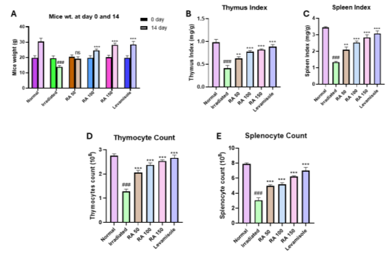
Fig. 1. RA effect on (A) mice weight, (B, C) thymic & splenic index, (D) thymocyte count, (E) splenocyte count. Normal, Irradiated, RA (50, 100, 150) mg/ kg, and levamisole 2.5 mg/kg (n = 6). Statistical analysis was performed using GraphPad software. Each group presents data as mean ± SD. #P ≤ 0.05, ##P ≤ 0.01, ###P ≤ 0.001 versus normal. *P ≤ 0.05, **P ≤ 0.01, ***P ≤ 0.001, ns: not significant, versus the irradiated group. RA: Rosmarinic acid
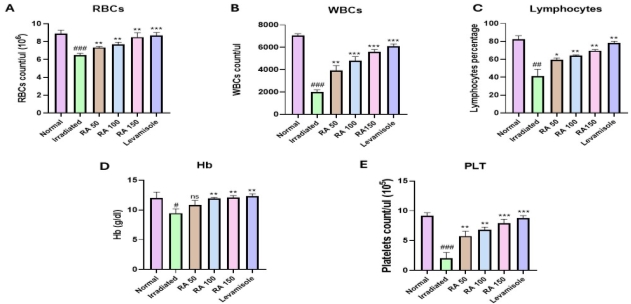
Fig. 2. Effects of RA on (A) RBCs, (B) WBCs, (C) lymphocytes percentage, (D) Hb, and (E) PLT. Normal, irradiated, RA (50, 100, 150) mg/ kg, and levamisole 2.5 mg/kg (n = 6). Statistical analysis was performed using GraphPad software. Each group presents data as means ± SD. #P ≤ 0.05, ##P ≤ 0.01, ###P ≤ 0.001 versus normal. *P ≤ 0.05, **P ≤ 0.01, ***P ≤ 0.001, ns: not significant, versus the irradiated group. RA: Rosmarinic acid; RBCs: Red blood cells; WBCs: White blood cells; Hb: Hemoglobin; PLT: platelets.
Effect of RA on Oxidant/Antioxidant Status: The effect of three different doses of RA on oxidant/antioxidant status in thymus and spleen after 5 Gy 137Cs γ-irradiation is shown in Figures 3 & 4, respectively.
As compared to irradiated groups, administration of RA significantly raised the level of SOD. In the normal group SOD activity was (0.303 ± 0.032IU/µL); in irradiated group, it decreased to 0.070 ± 0.020 IU/µL, while the high dose of RA increased this level to 0.253 ± 0.015 IU/µL (p ≤ 0.001. Fig. 3A).
The CAT activity of the normal group was 1.140 ± 0.052 IU/µL, which was lower in irradiated group 0.523 ± 0.076 IU/µL. RA increased this level to 0.885 ± 0.065 IU/µL (p ≤ 0.01) (Fig. 3C).
GPx increase was 74.6% in the higher dose (p ≤ 0.001) than in the irradiated group (Fig. 3D).
The MDA activity in thymus tissue of irradiated groups increased to 4.7050 ± 0.2150 µg/mg. In RA-treated groups, MDA level was reduced to 2.5817 ± 0.3325 µg/mg in the high dose (150mg/kg) group (Fig. 3B).
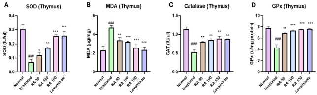
Fig. 3. Effects of RA on thymic (A) SOD, (B) MDA, (C) CAT, and (D) GPx. Normal, irradiated, RA (50, 100, 150) mg/ kg, and levamisole 2.5 mg/kg (n = 6). Statistical analysis was performed using GraphPad software. Each group presents data as means ± SD. #P ≤ 0.05, ##P ≤ 0.01, ###P ≤ 0.001 versus normal. *P ≤ 0.05, **P ≤ 0.01, ***P ≤ 0.001, ns: not significant, versus the irradiated group. RA: Rosmarinic acid; SOD: Superoxide dismutase; MDA: Malondialdehyde; CAT: Catalase; GPx: Glutathione peroxidase.
In the spleen, administration of RA significantly raised the levels of SOD p ≤ 0.001, GPx p ≤ 0.001, and CAT p ≤ 0.01, as compared to those in the untreated irradiated groups (Fig. 4A, C, & D).
SOD value of the normal group was 0.2067 ± 0.0031IU/µL, which decreased to 0.0633 ± 0.0058 IU/µL in the irradiated group. Treatment with high dose of RA increased SOD level to 0.1833 ± 0.0058 IU/µL, which was close to that in the levamisole treated group 0.1817 ± 0.0126 IU/µL.
The MDA activity in the spleen tissue of rosmarinic acid-treated groups declined to 2.868 ± 0.061 µg/mg in comparison to 4.693 ± 0.231 µg/mg (p ≤ 0.001) in the irradiated group. There was a trend towards improvement with RA treatment (Fig. 4B).
CAT activity of the normal group in spleen tissue was 1.087±0.132 IU/µL which decreased to (0.477±0.031 IU/µL) in irradiated group. RA dose at 150mg/kg increased this level to (0.882±0.076 IU/µL).
GPx activity was 9.057 ± 0.370(µ/mg of protein) in the normal group. In the irradiated group, this decreased to 5.167 ± 0.252 (µ/mg of protein) and with RA high dose, it increased to 6.617±0.246(µ/mg of protein).
Histopathology of the Thymus Gland and Spleen: The thymus of the normal group exhibited a distinct cortex with dark-staining cells adjacent to the medulla, paler-stained (Fig. 5A). The thymus of irradiated mice exhibited remarkable alterations in cellular density. The demarcation between the cortex and medulla looked less defined (Figure 6 5B). There were fewer degenerative changes in the thymus tissue of the RA-treated group as compared to that in the irradiated group (Table 2) (Fig. 5 C, D, E, F). The thymus tissues from the RA-treated group also showed a rise in lymphocytes (Fig. 5D).
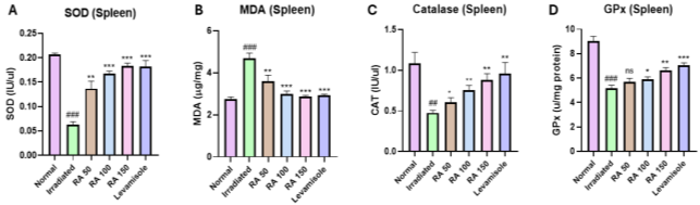
Fig. 4. Effects of RA on splenic (A) SOD, (B) MDA, (C) CAT, and (D) GPx. Normal, irradiated, RA (50,100, 150) mg/ kg, and levamisole 2.5 mg/kg (n = 6). Statistical analysis was performed using GraphPad software. Each group presents data as means ± SD. #P ≤ 0.05, ##P ≤ 0.01, ###P ≤ 0.001 versus normal. *P ≤ 0.05, **P ≤ 0.01, ***P ≤ 0.001, ns: not significant, versus the irradiated group. RA: Rosmarinic acid; SOD: Superoxide dismutase; MDA: Malondialdehyde; CAT: Catalase; GPx: Glutathione peroxidase.
Table 2. Thymus Histopathological Grading
|
Histopathological feature
|
Normal
|
Irradiated
|
RA 50 mg/kg
|
RA 100 mg/kg
|
RA 150 mg/kg
|
Levamisole
|
|
Lymphocyte depletion
|
0
|
3
|
2
|
1
|
1
|
0
|
|
Cortex changes
|
0
|
3
|
2
|
1
|
1
|
0
|
|
Medulla changes
|
0
|
2
|
2
|
1
|
0
|
0
|
|
Alteration in cellular density
|
0
|
3
|
2
|
1
|
1
|
0
|
|
Tissue degeneration
|
0
|
3
|
2
|
1
|
0
|
0
|
Scoring key: 0 = normal/absent, 1 = mild, 2 = moderate, 3 = severe
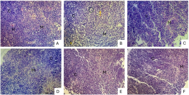
Fig. 5. Histological alterations in mouse thymus subjected to H&E staining. (A) Normal; (B) irradiated; (C) RA 50 mg/kg; (D) RA 100 mg/kg; (E) RA 150 mg/kg; (F) levamisole 2.5 mg/kg. Hassall's corpuscles (H), reticular cells (arrowhead), medulla (M), lymphocytes (arrow), cortex (C), and cell depletion (circle). Scale bar: (10 x). RA: Rosmarinic acid. H&E: Hematoxylin and Eosin stain.
The sections of splenic tissues of the normal group exhibited regular red and white pulp architecture, as well as thin splenic capsules. Spleen sections of irradiated mice exhibited disorganized splenic structure, marginally thick capsules, blood congestion, hemorrhage, and additional fibrous trabeculae. Whereas sections of splenic tissue of groups that were subjected to 5 Gy 137Cs γ-irradiation and various doses of RA ameliorated histological architecture to a different extent. Table 3. (Fig. 6).
Table 3. Spleen Histopathological Grading
|
Histopathological feature
|
Normal
|
Irradiated
|
RA 50 mg/kg
|
RA 100 mg/kg
|
RA 150 mg/kg
|
Levamisole
|
|
Splenic architectural disorganization
|
0
|
3
|
2
|
1
|
0
|
0
|
|
Red and white pulp damage
|
0
|
3
|
2
|
1
|
1
|
1
|
|
Capsule thickening
|
0
|
2
|
1
|
1
|
0
|
1
|
|
Blood congestion
|
0
|
3
|
2
|
1
|
1
|
1
|
|
Hemorrhage
|
0
|
2
|
1
|
1
|
0
|
0
|
|
Fibrous trabeculae
|
0
|
2
|
1
|
1
|
0
|
1
|
Scoring key: 0 = normal/absent, 1 = mild, 2 = moderate, 3 = severe
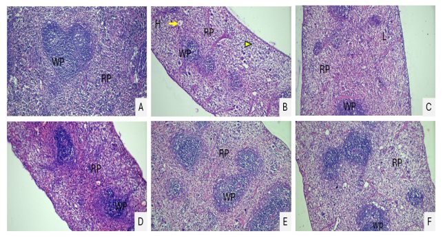
Fig. 6. Histopathological changes shown in splenic sections via H&E staining. (A) Normal; (B) irradiated; (C) RA 50 mg/kg; (D) RA 100 mg/kg; (E) RA 150 mg/kg; (F) levamisole 2.5 mg/kg. Cell depletion (circle), red pulps (RP), lymphocytes (arrowhead), white pulps (WP), Blood congestion (arrow), hemorrhage (H). (10 x). RA: Rosmarinic acid. H&E: Hematoxylin and Eosin stain.
Effect of RA on the Relative Expression Levels of IL-6, IL-7, IL-15, and FoxN1 in the Thymus: To further reveal the recovery effect of RA on thymic injury, we measured the relative mRNA expression of FoxN1 and thymic cytokines. Fig. 8A shows that relative fold expression of IL-7 decreased in the irradiated group, which was upregulated by all three doses of RA. Fig. 7B presented the data of IL-6 relative fold increase; high dose showed more increase in expression level. Fig. 7 C and D showed that levels of IL-15 and FoxN1 were inhabited in the irradiated group when compared to their levels in the normal group, RA doses upregulated their level. Fig. 7E is a visual image of gel bands. Relative mRNA expression of these genes increased with RA in the statistics of the irradiated group. After RA treatment at a (150mg/kg) dose, the expression level significantly improved (p ≤ 0.05).
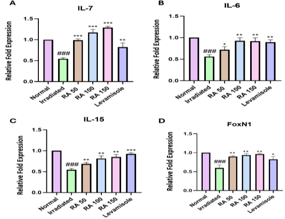
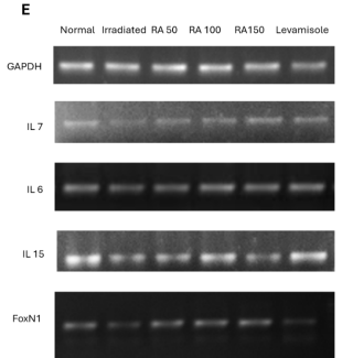
Fig. 7. PCR of (A) IL-7, (B) IL-6, (C) IL-15, (D) FoxN1, and (E) gel images. Normal, Irradiated; RA (50,100, 150) mg/ kg, and levamisole 2.5 mg/kg (n = 6). Statistical analysis was performed using GraphPad software. Each group presents data as means ± SD (n = 6). #P ≤ 0.05, ##P ≤ 0.01, ###P ≤ 0.001 versus normal. *P ≤ 0.05, **P ≤ 0.01, ***P ≤ 0.001, ns: not significant, versus the irradiated group. RA: Rosmarinic acid, IL: Interleukin.
DISCUSSION
Rosmarinic acid, a phenolic acid isolated from Rosmarinus officinalis, possesses anti-inflammatory, neuroprotective, antimicrobial, antioxidant, and diuretic properties (Ghaffari et al., 2014; Usha et al., 2014; Adomako-Bonsu et al., 2017; Nadeem et al., 2019; Moser et al., 2020). Given its documented antioxidant and anti-inflammatory properties, RA may counteract radiation-induced oxidative and inflammatory injury in lymphoid tissues. In the current research, we assessed the immunomodulatory effects of RA against radiation-induced injury on the spleen and thymus in mice. The results achieved in the current study were comparable to those observed in a standard positive-control group treated with levamisole, indicating the immunomodulatory effects of RA against radiation-induced injury.
In the present study, whole-body 5 Gy 137Cs γ-irradiation caused a considerable drop in the RBCs, WBCs, Hb, PLT, and lymphocyte. Similar findings were obtained by Begum et al, (2012), who observed a significant drop in the counts of RBCs and WBCs in mice exposed to 5 Gy 137Cs γ-irradiation. This phenomenon may be ascribed to the destruction and depletion of mature circulating blood cells due to hemorrhagic events or leakage via capillary membranes, coupled with a reduced cellular production (El-Desouky et al., 2017). Conversely, mice treated with RA displayed significant improvements in RBCs, WBCs, Hb, PLT, and lymphocyte counts.
The damage induced by radiation is principally attributed to the generation of free radicals, which substantially contribute to the biological effects of radiation and exerts deleterious effects on tissues and living organisms (Buonanno et al., 2011). During whole-body irradiation, the excessive generation of free radicals may overwhelm the antioxidant defense system, rendering it unable to neutralize them effectively. Antioxidants are effective scavengers of free radicals and are therefore considered promising candidates for the development of immunomodulators (Khan et al., 2015). Prior investigations have shown that RA serves as an efficacious antioxidant, effectively scavenging various free radicals, as the polyphenols facilitate the transfer of electrons, thereby promoting the antioxidant defense mechanisms (Ghali et al., 2024). These findings provided the rationale for investigating the modulatory effect of RA against radiation-induced damage in the thymus gland and spleen. The ability of RA to mitigate radiation-induced oxidative stress was assessed by measuring MDA, GPx, CAT, and SOD levels in the splenic and thymic tissue. These findings are consistent with those of Li et al. (2016), who reported a reduction in antioxidant enzyme activity and increased MDA levels in mice subjected to radiation. The present findings are also consistent with those of Fathy and Abdelkader (2021), who observed that an insecticide increased MDA activity and reduced antioxidant enzyme action in the thymic tissue of rats, while resveratrol, a polyphenolic compound, was able to reverse these alterations.
Ionizing radiation has the potential to induce mutations, chromosomal abnormalities, and apoptosis in cells (Mladenova et al., 2022). Typically, the tissues that exhibit the highest sensitivity to radiation are those characterized by rapid cellular turnover, such as the hematopoietic, gastrointestinal, and reproductive systems. Notably, immune cells present an exception to this generalization, as they display marked radiosensitivity despite their comparatively slow turnover rates (Talapko et al., 2024). Prior investigations have demonstrated that the thymus is particularly vulnerable to total-body irradiation, with sublethal doses causing both atrophy as well as structural alterations (Park et al., 2023; Duan et al., 2017). Following a 14 days regimen of sublethal-dose total body 5 Gy 137Cs γ-irradiation, we observed a statistically significant reduction in body weight, thymus weight, and thymocyte counts when compared with those in the control groups, potentially reflecting a decline in immune-cell populations.
Thymic atrophy was further characterized through histological examination of thymic tissue, which revealed a decrease in lymphocyte populations, the infiltration of macrophages. RA administration not only enhanced total body weight, thymic weight, and the thymic index but also facilitated the restoration of thymic tissue architecture with greater improvement generally observed at higher dose. Similarly, 5 Gy 137Cs γ-irradiation significantly reduced the spleen index, splenocyte count, and disrupted splenic histoarchitecture, while RA treatment markedly attenuated these alterations. These findings are consistent with the findings of prior reports. (Mahmoud et al., 2021).
Radiation-induced thymic damage resulted in acute involution, which was ameliorated by RA in the present work (Fig. 8). Under pathological conditions, the regeneration and functional restoration of TECs are essential for the re-establishment of an immunocompetent and self-tolerant T-cell repertoire (Hubscher et al., 2024). In our present research, we elucidated the effects of RA on the expression of immunoregulatory genes that are pivotal in T-cell differentiation, proliferation, and survival through their interaction with thymocytes. FoxN1 serves as a principal regulator of TECs (Li et al., 2023). IL-6 is implicated in the facilitation of an immunological response by enhancing the synthesis of antibodies (Tanaka et al., 2014; Saini et al., 2020). Given the pleiotropic role of IL-6 in inflammatory and immunoregulatory processes, its altered expression may reflect context-dependent changes in the immune response following irradiation. The finding are consistent with our previously published study (Mughal et al., 2024). IL-7 is a cytokine predominantly produced by TECs and fundamental component for the proliferation and survival of thymocytes (Bhalla et al., 2022). Furthermore, IL-7 shows a substantial role in stimulating the proliferation of double-negative thymocytes and in overall T-cell development (Gao et al., 2015). IL-15 is a potent immunostimulatory cytokine that facilitates the development of T cells. Previous investigations have demonstrated its pronounced effects on NK cell replies and in the activation of CD8+ T cells (Pilipow et al., 2015). The results from our current study revealed a reduction in the expression of the above-mentioned genes in the irradiated group, whereas RA treatment at various doses significantly improved their expression.
Altogether, the findings of the present study suggest that rosmarinic acid promotes the recovery of radiation-damaged thymic and splenic architecture and cellularity, thereby potentially ameliorating the deleterious effects of ionizing radiation.
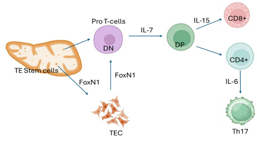
Fig. 8. Hypothetical model illustrates the proposed protective effect of rosmarinic acid against 5 Gy 137Cs γ-irradiation -induced ATI in mice.
Conclusions: The findings obtained in the present study suggested that RA attenuate 137Cs γ-radiation-induced injury in the thymus and spleen. RA ameliorated radiation-associated alterations in hematological parameters, body weight, thymocyte count, splenocyte counts, and thymic and splenic indices, and improved the radiation-induced histopathological changes in both lymphoid organs. Moreover, RA decreased radiation-induced oxidative stress, as indicated by reduced malondialdehyde levels and enhanced antioxidant enzyme activities in thymus and spleen. RA also modulated the radiation-induced expression of thymic IL-6, IL-7, IL-15, and FoxN1. Taken together, these findings proposed that RA may attenuate the harmful effects of radiation by decreasing oxidative stress, preserving lymphoid tissue architecture and cellularity, and modulating thymic gene expression. So, RA may have potential as a natural immunomodulatory agent against radiation-induced lymphoid injury.
Study Limitations: Numerous limitations of the present study should be acknowledged. First, the study was performed in one type of animal species and over 14 days of observation, which limits the long-term and chronic effect of RA. Second, survival and functional immune responses for long duration were not evaluated. Third, gene expression was analyzed without protein-level validation, and the findings were based primarily on mRNA analysis. Future studies can be performed with longer follow-up periods, functional immune assessments, survival analysis, and protein-level validation.
Acknowledgments: The authors are thankful to the host institute for providing the research environment.
Authors’ Contribution: A.R.: Investigation, validation, methodology, data curation, formal analysis, and initial drafting of the manuscript. R.M.: Visualization and proofreading of the manuscript. K.R.: Supervision, project administration, and validation. M.H.H.B.A.: Visualization, review, and proofreading of the manuscript. Z.U.: Conceptualization and proofreading of the manuscript. N.S.: Visualization, review, and proofreading of the manuscript. I.K.: Visualization, Resource, and proofreading of the manuscript. M.I.: Conceptualization, Supervision, project administration, conceptualization, methodology, formal analysis, data curation, validation, and review and final editing of the manuscript.
REFERENCES
Adomako-Bonsu, A.G., S.L. Chan, M. Pratten and J.R. Fry (2017). Antioxidant activity of rosmarinic acid and its principal metabolites in chemical and cellular systems: importance of physico-chemical characteristics. Toxicol. Vitro. 40(1): 248-255. https://doi.org/10.1016/j.tiv.2017.01.016.
Barbouti, A., P.V. Vasileiou, K. Evangelou, K.G. Vlasis, A. Papoudou-Bai, V.G. Gorgoulis and P. Kanavaros (2020). Implications of oxidative stress and cellular senescence in age‐related thymus involution. Oxid. Med. Cell. Longev. 2020(1): 7986071-7986085. https://doi.org/10.1155/2020/7986071.
Begum, N., N.R. Prasad and K. Thayalan (2012). Apigenin protects gamma-radiation induced oxidative stress, hematological changes and animal survival in whole body irradiated Swiss albino mice. Int. J. Nutr. Pharmacol. Neurol. Dis. 2(1): 45-52. https://doi.org/10.4103/2231-0738.93134.
Bhalla, P., D.M. Su and N.S. Vanoers (2022). Thymus functionality needs more than a few TECs. Front. Immunol. 13(1): 864777-864791. https://doi.org/10.3389/fimmu.2022.864777.
Bhangale, J.O. and S.R. Acharya (2016). Anti-Parkinson activity of petroleum ether extract of Ficus religiosa (L.) leaves. Adv. Pharmacol. Pharm. Sci. 2016(1): 9436106-9436115. https://doi.org/10.1155/2016/9436106.
Boonyarikpunchai, W., S. Sukrong and P. Towiwat (2014). Antinociceptive and anti-inflammatory effects of rosmarinic acid isolated from Thunbergia laurifolia Lindl. Pharmacol. Biochem. Behav. 124(1): 67-73. https://doi.org/10.1016/j.pbb.2014.05.004.
Buonanno, M., S.M. De Toledo, D. Pain and E.I. Azzam (2011). Long-term consequences of radiation-induced bystander effects depend on radiation quality and dose and correlate with oxidative stress. Radiat. Res. 175(4): 405-415. https://doi.org/10.1667/RR2461.1.
Cassatt, D.R., T.A. Winters and M. PrabhuDas (2023). Immune dysfunction from radiation exposure. Radiat. Res. 200(4): 389-395. https://doi.org/10.1667/RADE-22-00004.1.
Comelekoglu, U., S. Yalin, S. Bagis, O. Ogenler, N.O. Sahin, A. Yildiz, B. Coskun, R. Hatungil and A. Turac (2007). Low-exposure cadmium is more toxic on osteoporotic rat femoral bone: mechanical, biochemical, and histopathological evaluation. Ecotoxicol. Environ. Saf. 66(2): 267-271. https://doi.org/10.1016/j.ecoenv.2006.01.006.
Duan, Y., X. Yao, J. Zhu, Y. Li, J. Zhang, X. Zhou, Y. Qiao, M. Yang and X. Li (2017). Effects of yak-activated protein on hematopoiesis and related cytokines in radiation-induced injury in mice. Exp. Ther. Med. 14(6): 5297-5304. https://doi.org/10.3892/etm.2017.5256.
El-Desouky, W., A. Hanafi and M.M. Abbas (2017). Radioprotective effect of green tea and grape seed extracts mixture on gamma irradiation induced immune suppression in male albino rats. Int. J. Radiat. Biol. 93(4): 433-439. https://doi.org/10.1080/09553002.2016.1254834.
Fan, H., S. Zhang, N. Li, P. Fan, X. Hu, K. Liang, X. Chang and Y. Wu (2020). Stable expression ratios of five pyroptosis-inducing cytokines in the spleen and thymus of mice showed potential immune regulation at the organ level. Lupus. 29(3): 290-302. https://doi.org/10.1177/0961203320903802.
Fathy, S.M. and I.Y. Abdelkader (2021). Effect of resveratrol on the inflammatory status and oxidative stress in thymus gland and spleen of sulfoxaflor‐treated rats. Environ. Toxicol. 36(7): 1326-1337. https://doi.org/10.1002/tox.23129.
Gao, J., L. Zhao, Y.Y. Wan and B. Zhu (2015). Mechanism of action of IL-7 and its potential applications and limitations in cancer immunotherapy. Int. J. Mol. Sci. 16(5): 10267-10280. https://doi.org/10.3390/ijms160510267.
Gayathri, V., V.V. Asha, J.A. John and A. Subramoniam (2011). Protection of immunocompromised mice from fungal infection with a thymus growth-stimulatory component from Selaginella involvens, a fern. Immunopharmacol. Immunotoxicol. 33(2): 351-359. https://doi.org/10.3109/08923973.2010.518617.
Ghaffari, H., M. Venkataramana, B.J. Ghassam, S.C. Nayaka, A. Nataraju, N.P. Geetha and H.S. Prakash (2014). Rosmarinic acid mediated neuroprotective effects against H2O2-induced neuronal cell damage in N2A cells. Life Sci. 113(2): 7-13. https://doi.org/10.1016/j.lfs.2014.07.010.
Ghali, E.H.K., S.K. Sandopu, D.K. Maurya and B. Meriga (2024). Insights into the radioprotective efficacy of Pterocarpus santalinus L. aqueous extract. Fitoterapia. 176(1): 105986-105998. https://doi.org/10.1016/j.fitote.2024.105986.
Gong, Y., J. Wu and S.T. Li (2015). Immuno-enhancement effects of Lycium ruthenicum Murr. polysaccharide on cyclophosphamide-induced immunosuppression in mice. Int. J. Clin. Exp. Med. 8(11): 20631-20638. PMID: 26884983.
Granadier, D., D. Acenas and J.A. Dudakov (2025). Endogenous thymic regeneration: restoring T cell production following injury. Nat. Rev. Immunol. 25(6): 407-424. https://doi.org/10.1038/s41577-024-01119-0.
Guan, H., W. Luo, B. Bao, Y. Cao, F. Cheng, S. Yu, Q. Fan, L. Zhang, Q. Wu and M. Shan (2022). A comprehensive review of rosmarinic acid: from phytochemistry to pharmacology and its new insight. Mol. 27(10): 3292-3320. https://doi.org/10.3390/molecules27103292.
Horie, K., K. Namiki, K. Kinoshita, M. Miyauchi, T. Ishikawa, M. Hayama, Y. Maruyama, N. Hagiwara, T. Miyao, S. Murata and T.J. Kobayashi (2023). Acute irradiation causes a long-term disturbance in the heterogeneity and gene expression profile of medullary thymic epithelial cells. Front. Immunol. 14(1): 1186154-1186167. https://doi.org/10.3389/fimmu.2023.1186154.
Hubscher, T., L.F. Lorenzo-Martín, T. Barthlott, L. Tillard, J.J. Langer, P. Rouse, C.C. Blackburn, G. Hollander and M.P. Lutolf (2024). Thymic epithelial organoids mediate T-cell development. Dev. 151(17): 202853-202865. https://doi.org/10.1242/dev.202853.
Hwang, Y.P., G.H. Lee, T.H. Pham, M.Y. Kim, C.Y. Kim, S.Y. Lee, E.H. Han, C.Y. Choi, S.D. Hwang, S. Ahn and H.G. Jeong (2022). Immune-enhancing effect of submerged culture of Ceriporia lacerata mycelia on cyclophosphamide-induced immunosuppressed mice and the underlying mechanisms in macrophages. Int. J. Mol. Sci. 23(2): 597-605. https://doi.org/10.3390/ijms23020597.
Khan, S., J.S. Adhikari, M.A. Rizvi and N.K. Chaudhury (2015). Radioprotective potential of melatonin against 60Co γ-ray-induced testicular injury in male C57BL/6 mice. J. Biomed. Sci. 22(1): 61-76. https://doi.org/10.1186/s12929-015-0156-9.
Kinsella, S. and J.A. Dudakov (2020). When the damage is done: injury and repair in thymus function. Front. Immunol. 11(1): 1745-1757. https://doi.org/10.3389/fimmu.2020.01745.
Kuo, J.F., H.Y. Wu, C.W. Tung, W.H. Huang, C.S. Lin and C.C. Wang (2024). Induction of thymus atrophy and disruption of thymocyte development by fipronil through dysregulation of IL-7-associated genes. Chem. Res. Toxicol. 37(9): 1488-1500. https://doi.org/10.1021/acs.chemrestox.4c00060.
Lee, D.Y., W.H. Song, Y.S. Lim, C. Lee, L. Rajbongshi, S.Y. Hwang, B.S. Kim, D. Lee, Y.J. Song, H.G. Kim and S. Yoon (2023). Fish collagen peptides enhance thymopoietic gene expression, cell proliferation, thymocyte adherence, and cytoprotection in thymic epithelial cells via activation of the nuclear factor-κB pathway, leading to thymus regeneration after cyclophosphamide-induced injury. Mar. Drugs. 21(10): 531-564. https://doi.org/10.3390/md21100531.
Lewis, S.M., A. Williams and S.C. Eisenbarth (2019). Structure and function of the immune system in the spleen. Sci. Immunol. 4(33): 6085-6098. https://doi.org/10.1126/sciimmunol.aau6085.
Li, J., L.P. Wachsmuth, S. Xiao, B.G. Condie and N.R. Manley (2023). Foxn1 overexpression promotes thymic epithelial progenitor cell proliferation and mTEC maintenance but does not prevent thymic involution. Dev. 150(8): 200995-201007. https://doi.org/10.1242/dev.200995.
Li, N., X. Shen, Y. Liu, J. Zhang, Y. He, Q. Liu, D. Jiang, J. Zong, J. Li, D. Hou and W. Chen (2016). Isolation, characterization, and radiation protection of Sipunculus nudus L. polysaccharide. Int. J. Biol. Macromol. 83(1): 288-296. https://doi.org/10.1016/j.ijbiomac.2015.11.071.
Li, Y., H. Li, C. Peng, G. Meng, Y. Lu, H. Liu, L. Cui, H. Zhou, Z. Xu, L. Sun and L. Liu (2024). Unraveling the spatial organization and development of human thymocytes through integration of spatial transcriptomics and single-cell multi-omics profiling. Nat. Commun. 15(1): 7784-7809. https://doi.org/10.1038/s41467-024-51767-y.
Liu, Z., Q. Liang, Y. Ren, C. Guo, X. Ge, L. Wang, Q. Cheng, P. Luo, Y. Zhang and X. Han (2023). Immunosenescence: molecular mechanisms and diseases. Signal. Transduct. Target. Ther. 8(1): 200-216. https://doi.org/10.1038/s41392-023-01451-2.
Mahmoud, H.S., A.A. Almallah, H.N. Gad EL-Hak, T.S. Aldayel, H.M. Abdelrazek and H.E. Khaled (2021). The effect of dietary supplementation with Nigella sativa (black seeds) mediates immunological function in male Wistar rats. Sci. Rep. 11(1): 7542-7555. https://doi.org/10.1038/s41598-021-86721-1.
Mladenova, V., E. Mladenov, M. Stuschke and G. Iliakis (2022). DNA damage clustering after ionizing radiation and consequences in the processing of chromatin breaks. Mol. 27(5): 1540-1558. https://doi.org/10.3390/molecules27051540.
Moser, J.C., C.C. Cechinel-Zanchett, L.N.B. Mariano, T. Boeing, L.M. da Silva and P. de Souza (2020). Diuretic, natriuretic and Ca2+-sparing effects induced by rosmarinic and caffeic acids in rats. Rev. Bras. Farmacogn. 30(4): 588-592. https://doi.org/10.1007/s43450-020-00075-9.
Mughal, K.S., M. Ikram, Z. Uddin, A. Rashid, U. Rashid, M. Khan, N. Zehra, U.S. Mughal, N. Shah and I. Amirzada (2024). Syringic acid improves cyclophosphamide-induced immunosuppression in a mouse model. Biochem. Biophys. Res. Commun. 734(1): 150777-150790. https://doi.org/10.1016/j.bbrc.2024.150777.
Nadeem, M., M. Imran, T. Aslam Gondal, A. Imran, M. Shahbaz, R. Muhammad Amir, M. Waseem Sajid, T. Batool Qaisrani, M. Atif, G. Hussain and B. Salehi (2019). Therapeutic potential of rosmarinic acid: a comprehensive review. Appl. Sci. 9(15): 3139-3162. https://doi.org/10.3390/app9153139.
Park, H.R., J.W. Kim, J.O. Lee, J.D. Ahn, M.C. Yang, K.M.I. Bashir, J.S. Choi and S.K. Ku (2023). Anti-mutagenic and immunomodulatory effects of Astragali radix extract on a cyclophosphamide-induced immunosuppressed mouse model. Appl. Sci. 13(5): 2959-2979. https://doi.org/10.3390/app13052959.
Pilipow, K., A. Roberto, M. Roederer, T.A. Waldmann, D. Mavilio and E. Lugli (2015). IL15 and T-cell stemness in T-cell–based cancer immunotherapy. Cancer Res. 75(24): 5187-5193. https://doi.org/10.1158/0008-5472.CAN-15-1498.
Saini, C., R.K. Srivastava, M. Tarique, S. Kurra, N. Khanna, V. Ramesh and A. Sharma (2020). Elevated IL-6R on CD4+ T cells promotes IL-6 driven Th17 cell responses in patients with T1R leprosy reactions. Sci. Rep. 10(1): 15143-15156. https://doi.org/10.1038/s41598-020-72148-7.
Saleem, U., Z. Chauhdary, Z. Raza, S. Shah, M.U. Rahman, P. Zaib and B. Ahmad (2020). Anti-parkinson’s activity of Tribulus terrestris via modulation of AChE, α-synuclein, TNF-α, and IL-1β. ACS Omega. 5(39): 25216-25227. https://doi.org/10.1021/acsomega.0c03375.
Saleem, U., S. Hira, F. Anwar, M.A. Shah, S. Bashir, R.S. Baty, R.H. Badr, R. Blundell, G.E.S. Batiha and B. Ahmad (2021). Pharmacological screening of Viola odorata L. for memory-enhancing effect via modulation of oxidative stress and inflammatory biomarkers. Front. Pharmacol. 12(1): 664832-664844. https://doi.org/10.3389/fphar.2021.664832.
Talapko, J., D. Talapko, D. Katalinić, I. Kotris, I. Erić, D. Belić, M. Vasilj Mihaljević, A. Vasilj, S. Eric, J. Flam and S. Bekic (2024). Health effects of ionizing radiation on the human body. Medicina. 60(4): 653-670. https://doi.org/10.3390/medicina60040653.
Tanaka, T., M. Narazaki and T. Kishimoto (2014). IL-6 in inflammation, immunity, and disease. Cold Spring Harb. Perspect. Biol. 6(10): 016295-016312. https://doi.org/10.1101/cshperspect.a016295.
Thapa, P. and D.L. Farber (2019). The role of the thymus in the immune response. Thorac. Surg. Clin. 29(2): 123-131. https://doi.org/10.1016/j.thorsurg.2018.12.001.
Usha, T., S.K. Middha, M. Bhattacharya, P. Lokesh and A.K. Goyal (2014). Rosmarinic acid, a new polyphenol from Baccaurea ramiflora Lour. leaf: a probable compound for its anti-inflammatory activity. Antioxidants. 3(4): 830-842. https://doi.org/10.3390/antiox3040830.
Velardi, E., J.J. Tsai and M.R. Van Den Brink (2021). T cell regeneration after immunological injury. Nat. Rev. Immunol. 21(5): 277-291. https://doi.org/10.1038/s41577-020-00457-z.
Wang, L. and X. Li (2019). Radioprotective effect of Hohenbuehelia serotina polysaccharides through mediation of ER apoptosis pathway in vivo. Int. J. Biol. Macromol. 127(1): 18-26. https://doi.org/10.1016/j.ijbiomac.2018.12.267.
Zhu, N., R. Liu, L.X. He, R.X. Mao, X.R. Liu, T. Zhang, Y.T. Hao, R. Fan, M.H. Xu and Y. Li (2019). Radioprotective effect of walnut oligopeptides against gamma radiation-induced splenocyte apoptosis and intestinal injury in mice. Mol. 24(8): 1582-1598. https://doi.org/10.3390/molecules24081582.